 Who moved my microcentrifuge tube? (and my PCR tubes)
 Follow Simport technological breakthrough in immunofluorescence: The StainTray
 Pasteur pipets or transfer pipets?
 Introducing High-Density Polyethylene (HDPE)
 General
 Applications
NEED HELP?
(450) 464-1723
1-800-464-1723
1-800-464-1723


Published :
01/20/2023 10:37:46
Categories :
General

Obviously, this article is not to explain this technique to cytotechnologists, who understand the details of this laboratory method. Instead, it is intended for our partners, distributors, and representatives to improve their understanding and the possibilities of these strangely shaped laboratory consumables.
Cyto- means this technique deals with the study of cells. However, this method is used only to prepare cells for examination, not the exam itself. Therefore, focusing on preparing a smear, a single layer (monolayer) of cells on a glass microscope slide.
Centrifugation means that this technique will use a centrifuge to achieve its goal. The centrifuge uses centrifugal force to separate elements in a liquid. Specifically, forcing large particles, like cells, from floating and directing them toward the glass slide.
The fluids we are referring to here could be any body fluid; urine, pleural, synovial cerebrospinal, or bronchial aspirates. Thus any liquid in which the clinician wants to examine the cells it contains if any.
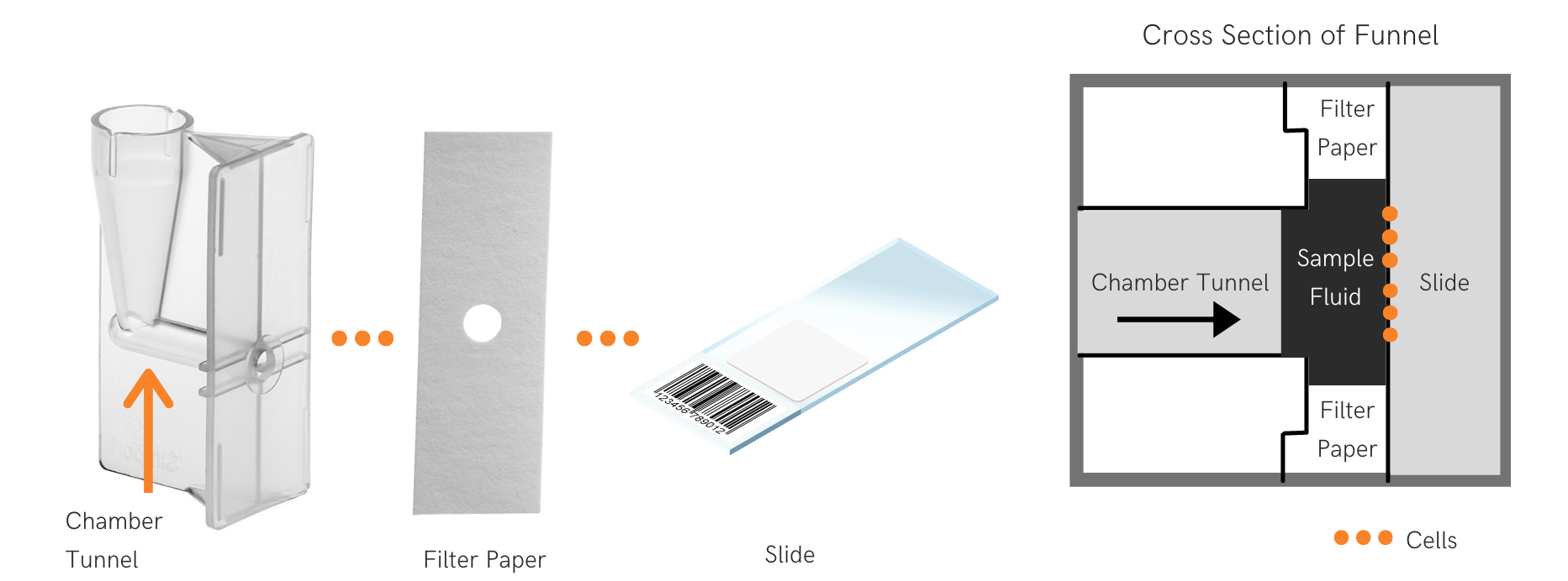
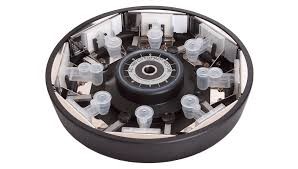
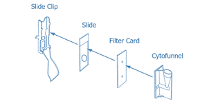
Here comes our cytofunnel. As the name describes, this item is a funnel equipped with a horizontal channel at its bottom, coupled to a flat vertical surface. The fluid is placed in the funnel once a microscope glass slide is installed by means of a metal clip or with an integrated disposable mechanism. Once ready, the cytofunnel is placed in the centrifuge and spun at a relatively low speed. Cells are fragile little things, destroyed by high speed and g forces. Moderate speeds are more than sufficient for this procedure.
During the centrifugation, here is what happens. The liquid will have filled the channel at the bottom of the funnel, creating a space through which cells can travel. And they do, pushed by the centrifugal force. They travel through the liquid until they reach the glass slide on which they deposited. The remaining liquid is either left there and decanted before dismantling the apparatus or, more frequently nowadays, absorbed by a filter placed strategically between the cytofunnel and the glass slide. Correctly performed, the result is a nice monolayer smear of cells on the glass slide with no residual liquid. The slide is then taken to the staining station and treated in order to become visible under a microscope to be studied.
There are variations. Clips are available as metallic making them reusable or integrated into the funnel and disposable. Filters may be separate or pre-attached to the cytofunnel. Funnels are offered in various sizes to accommodate the volume of tested fluid. Different shapes are available as well in order to adapt to the variety of centrifuge models. A denser brown filter is even offered that discharges less fiber for use with samples with very few cells like CSF!
Hopefully, this article helped you understand cytocentrifugation. All that remains is to discover Simport's great selection of high-quality cytofunnels for all laboratories wanting to provide clinicians with the highest-quality smears!
.png)